INTRODUCTION — Acute appendicitis is the most common general surgical problem encountered during pregnancy . The incidence ranges from 0.06 to 0.1 percent, or 1 in 1500 deliveries . However, pregnant women appear to be less likely to have appendicitis than age-matched, nonpregnant women.
CLINICAL MANIFESTATIONS AND DIAGNOSTIC EVALUATION
Clinical manifestations and diagnosis — The clinical manifestations and diagnostic evaluation of appendicitis in pregnancy are similar to those in nonpregnant individuals and are described in detail separately .
Certain points, however, apply to pregnant women:
• There is a slightly higher rate of appendicitis in the second trimester than in the first and third trimesters or postpartum . An infected appendix appears to be more likely to rupture during pregnancy, especially in the third trimester, possibly because of delay in diagnosis and intervention .
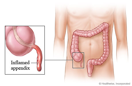
• Right lower quadrant pain is the most common symptom of appendicitis and should alert the physician caring for the pregnant patient to strongly consider this diagnosis. Although the location of the appendix migrates a few centimeters cephalad with the enlarging uterus , the most common symptom of appendicitis, ie, right lower quadrant pain, occurs close to McBurney's point in the vast majority of pregnant women, regardless of the stage of pregnancy.
Differential diagnosis — The differential diagnosis includes the causes of abdominal pain in nonpregnant individuals , as well as pregnancy-related causes of abdominal pain, such as round ligament syndrome, labor, abruption, ectopic pregnancy, and uterine rupture . The latter three diagnoses, in contrast to appendicitis, are often accompanied by uterine bleeding. In addition, abruption and uterine rupture are commonly associated with fetal heart rate abnormalities. Preeclampsia and HELLP syndrome can be associated with abdominal pain, but it is usually in the right upper quadrant or epigastrium.
The diagnosis of acute appendicitis in a laboring patient is especially difficult and requires a high index of suspicion. Labor can be associated with pain that may be lateralized, fever if chorioamnionitis is present, leukocytosis, and vomiting.
• The physiologic changes of pregnancy can confound the diagnosis. In particular, the normal pregnant woman's white blood cell count ranges from 6000 to 16,000 cell/mm3 in the first and second trimesters, and may rise as high as 20,000 to 30,000 cells/mm3 during labor . Thus, leukocytosis can be a normal finding in pregnant women. This was illustrated in a retrospective review of 66,993 consecutive deliveries which included 67 patients with a probable diagnosis of appendicitis . Patients with a confirmed diagnosis of appendicitis had a mean leukocyte count of 16,400 cells/mm3 versus 14,000 cells/mm3 for patients with a normal appendix.
• Indigestion, bowel irregularity, nausea/vomiting, and a sense of not feeling well are common symptoms of both appendicitis and normal pregnancy. In appendicitis, nausea and vomiting, if they occur, follow the onset of pain, whereas nausea and vomiting associated with pregnancy is not usually associated with pain.
• Pregnant women with pyuria may be treated for urinary tract infection and forgo further investigation, in which case the actual diagnosis of appendicitis may be delayed. It is important to remember the inflamed appendix is often in close proximity to the bladder and ureter; as a result, microscopic hematuria and pyuria are found in up to one-third of patients with acute appendicitis.
• Peritoneal findings may be less prominent than in nonpregnant women because the gravid uterus lifts and stretches the anterior abdominal wall away from the inflamed appendix . Since direct contact between the area of inflammation and parietal peritoneum is impeded, there is less muscle response or guarding. The gravid uterus may also inhibit contact between the omentum and the inflamed appendix.
Imaging — As in nonpregnant patients, diagnostic imaging should be performed in patients suspected of having appendicitis when the diagnosis is unclear after assessment of presenting complaints, physical examination, and laboratory results . Thus, virtually all pregnant women will have an imaging study.
Ultrasonography — The initial imaging modality of choice for diagnostic imaging of the appendix in pregnancy is graded compression ultrasonography . Ultrasound allows for visualization of the uterus, placenta and ovaries, and thus can be used to exclude some other causes of right lower quadrant pain. Appendicitis is diagnosed if a noncompressible blind ended tubular structure is visualized in the right lower quadrant with a maximal diameter greater than 6 mm . As a general rule, the main role of ultrasonography is to help confirm the diagnosis of suspected appendicitis; if a normal appendix is not visualized, appendicitis cannot be excluded.
A systematic review of this technique in individuals 14 years of age and older reported overall sensitivity of 86 percent (95% CI 83-88), specificity 81 percent (95% CI 78-84), positive likelihood ratio 5.8 (95% CI 3.5-9.5), and negative likelihood ratio 0.19 (95% CI 0.13-0.27) [19].
However, the gravid uterus can interfere with performance of this technique, particularly in the third trimester, leading to a high negative laparotomy rate when ultrasound results are inconclusive . There are no large series in pregnant women. In one small series, the appendix could not be visualized with ultrasound in 22 of 23 pregnant patients with suspected appendicitis .
Magnetic resonance imaging — Where readily available, magnetic resonance imaging (MRI) can be useful for the next step in diagnostic evaluation of cases with diagnostic uncertainty. MRI offers an attractive alternative to CT because it avoids exposure to ionizing radiation. Observational data suggest that MRI can accurately diagnose appendicitis during pregnancy .
• The largest study consisted of 148 consecutive pregnant women who underwent MRI because of clinically suspected appendicitis; 140 of these women also had an ultrasound examination . The appendix was imaged by MRI in 130 patients (88 percent), 14/14 with and 116/134 without appendicitis. The sensitivity, specificity, positive and negative predictive values of MRI for diagnosis of acute appendicitis were 100, 93, 61, and 100 percent, respectively.
• A meta-analysis reported similar findings, but the results are limited because the analysis included only four small studies of 12 to 51 patients who had normal or inconclusive ultrasonography, and these studies included only three to five patients with appendicitis . The authors calculated sensitivity 80 percent (95% CI 44-98) and specificity 99 percent (94 to 100).
MRI appears to be an excellent modality for excluding acute appendicitis in pregnant women who present with characteristic signs and symptoms and in whom ultrasound examination is inconclusive.
If there is going to be a prolonged wait before an MRI can be performed, the increasing risk of rupture over time should be considered and undue delays for imaging studies should be avoided.
Computed tomography — Computed tomography (CT) is generally widely available. The main findings of appendicitis on CT are right lower quadrant inflammation, an enlarged nonfilling tubular structure, and/or an appendicolith . The initial experience with helical computed tomography for the diagnosis of appendicitis in pregnancy appears promising, but data are limited to small case series . Modifications to the CT protocol limit estimated fetal radiation exposure to less than 300 mrad, well below doses known to cause adverse fetal effects, and do not limit diagnostic performance . Standard abdominal CT scanning with an oral preparation and intravenous contrast or a specialized appendiceal CT scanning protocol can be used. The relative advantages and disadvantages of the two protocols and what constitutes a positive study are described separately.
We suggest CT when clinical findings and ultrasound are inconclusive and magnetic resonance imaging is not available, given the proven diagnostic value of CT in nonpregnant individuals: overall sensitivity 94 percent (95% CI 91-95), specificity 95 percent (95% CI 93-96), positive likelihood ratio 13.3 (95% CI 9.9-17.9), and negative likelihood ratio 0.09 (95% CI 0.07-0.12) . In addition, a meta-analysis of three retrospective studies in pregnant women reported the sensitivity and specificity of CT in cases of normal/uncertain ultrasonography were: sensitivity 85.7 percent (95% CI 63.7-96) and specificity 97.4 percent (95% CI 86.2-99.9) . These studies included two to 49 patients with appendicitis.
MANAGEMENT APPROACH AND OUTCOME — Consultation with a general surgeon can be useful in women whose imaging studies suggest appendicitis. The decision to proceed to laparotomy should be based upon the clinical findings, diagnostic imaging results, and clinical judgment. Laboratory tests are not particularly useful other than to rule in an alternate diagnosis. Delaying intervention for more than 24 hours increases the risk of perforation , which occurs in 14 to 43 percent of such patients. The risk of fetal loss is also higher in patients in whom the appendix had perforated (36 versus 1.5 percent) or when there is generalized peritonitis or a peritoneal abscess (fetal loss: 6 versus 2 percent; early delivery: 11 versus 4 percent ). Given the diagnostic difficulties and significant risk of fetal mortality with perforation, a higher negative laparotomy rate (20 to 35 percent) compared to nonpregnant women has generally been considered to be acceptable. Aggressive use of radiologic imaging, especially MR and CT scanning, may reduce the incidence of negative appendectomy.
Maternal morbidity following appendectomy is low except in patients in whom the appendix has perforated. In contrast, pregnancy related complications are frequent, particularly when surgery was performed in the first or second trimester. This was illustrated in a series of 56 women who underwent appendectomy in various trimesters . Spontaneous abortion was observed in 4 of 12 patients (33 percent) who underwent appendectomy in the first trimester while 4 of 28 (14 percent) patients operated on in the second trimester delivered prematurely. No pregnancy complications were observed in women who underwent appendectomy in the third trimester.
Cesarean delivery is rarely indicated at the time of appendectomy. Moreover, the risk of dehiscence during labor and vaginal delivery should not be increased when the fascia has been appropriately reapproximated .
The long-term prognosis for women who undergo appendectomy during pregnancy seems to be good. Such women do not appear to be at increased risk for infertility or other complications.
Perforated appendix — The management of appendiceal perforation depends on the nature of the perforation. A free perforation can cause intraperitoneal dissemination of pus and fecal material. These patients are typically quite ill and may be septic; as discussed above, they are at increased risk of preterm labor and delivery and fetal loss . Urgent laparotomy is necessary with appendectomy and irrigation and drainage of the peritoneal cavity.
Nonpregnant patients who present with a long duration of symptoms (more than five days) and have findings of a contained perforation can be treated initially with antibiotics, intravenous fluids, bowel rest, and close monitoring. These patients will often have a palpable mass on physical examination and imaging may reveal a phlegmon or abscess. Fortunately, many of these patients will respond to nonoperative management since the appendiceal process has already been "walled-off."
Conservative treatment is a good option since immediate surgery in patients with a long duration of symptoms and phlegmon formation is associated with increased morbidity due to dense adhesions and inflammation. Under these circumstances, appendectomy often requires extensive dissection and may lead to injury of adjacent structures. Complications such as a postoperative abscess, or enterocutaneous fistula may ensue, necessitating an ileocolectomy or cecostomy. Because of these potential complications, a non-operative approach can be considered if the patient is not ill-appearing.
Although there is good evidence to support this approach in nonpregnant individuals, there is only limited evidence in pregnant women. In a single report including only two patients, antibiotic therapy (ampicillin, gentamicin, and clindamycin), intravenous fluids, and bowel rest was associated with improvement in symptoms over two to three days. In one patient, interval appendectomy was performed two months post-vaginal delivery. In the other patient, appendectomy was performed at cesarean delivery because of breech presentation with preterm labor; this patient had an appendiceal phlegmon that had been treated conservatively seven weeks earlier, but with recurrence of acute appendiceal inflammation. In both cases, treatment with antenatal glucocorticoids to induce fetal lung maturation and tocolytics were avoided due to concerns of suppressing clinical manifestations of worsening infection and delaying delivery if intraamniotic infection was also present. Until further experience of non-operative management of ruptured, but contained, appendicitis during pregnancy is available, such patients should be followed closely in the hospital to monitor for maternal sepsis and preterm labor.
SURGICAL APPROACH — The intraoperative management of pregnant women and monitoring of the fetus are reviewed separately.
When the diagnosis is relatively certain, we suggest performing appendectomy through a transverse incision at McBurney's point, or more commonly, over the point of maximal tenderness . When the diagnosis is less certain, we suggest a lower midline vertical incision since it permits adequate exposure of the abdomen for diagnosis and treatment of surgical conditions that mimic appendicitis. A vertical incision can also be used for a cesarean delivery, if subsequently required for the usual obstetric indications.
There have been several case reports and small case series on the use of laparoscopic appendectomy in pregnancy that suggested such procedures can be performed successfully during all trimesters and with few complications . Although long-term data on the safety and efficacy of laparoscopic appendectomy during pregnancy are limited, one systematic review noted a higher rate of fetal loss with this approach than with open appendectomy, but these data were from retrospective series .
The decision to proceed with a laparoscopic approach should take into consideration the skill and experience of the surgeon, as well as clinical factors such as the size of the gravid uterus. Suggestions for modification of laparoscopic technique during pregnancy include slight left lateral positioning of the patient during the second half of pregnancy, avoiding the use of any cervical instruments, consideration of open entry techniques or placement of trocars under direct visualization, and limiting intra-abdominal pressure to less than 12 mmHg .
SUMMARY AND RECOMMENDATIONS
• Acute appendicitis is the most common general surgical problem encountered during pregnancy.
• The clinical manifestations and diagnosis of appendicitis in pregnancy are similar to those in nonpregnant individuals; however, the following points should be noted.
- Right lower quadrant pain is the most common symptom and occurs within a few centimeters of McBurney's point in the vast majority of pregnant women, regardless of the stage of pregnancy.
- The normal pregnant woman's white blood cell count ranges from 6000 to 16,000 cell/mm3 in the first and second trimesters, and may rise as high as 20,000 to 30,000 cells/mm3 during labor so leukocytosis may or may not be a sign of appendicitis.
- Indigestion, bowel irregularity, nausea/vomiting, and a sense of not feeling well are common symptoms of both appendicitis and normal pregnancy. In appendicitis, nausea and vomiting, if they occur, follow the onset of pain, whereas nausea and vomiting associated with pregnancy is not usually associated with pain.
• We suggest graded compression ultrasonography in pregnant patients suspected of having appendicitis. If ultrasonography suggests appendicitis, a general surgeon should be consulted.
• If clinical findings and ultrasound are inconclusive, or in centers where experience with sonographic examination of the appendix is limited, we suggest magnetic resonance imaging, where available, because it avoids fetal exposure to ionizing radiation and performs well in diagnosis of lower abdominal/pelvic disorders. If magnetic resonance imaging suggests appendicitis, a general surgeon should be consulted.
We suggest computed tomography when magnetic resonance imaging is not available, given its proven value in nonpregnant individuals. (See 'Computed tomography' above.)
• The decision to proceed to laparotomy should be based upon the clinical findings, diagnostic imaging results, and clinical judgment. Laboratory tests are not particularly useful other than to rule in an alternate diagnosis. Delaying intervention for more than 24 hours increases the risk of perforation.
• The risk of dehiscence during labor and vaginal delivery should not be increased when the fascia has been appropriately reapproximated.
• When the diagnosis is relatively certain, we suggest performing appendectomy through a transverse incision over the point of maximal tenderness (Grade 2C). When the diagnosis is less certain, we suggest a lower midline vertical incision (Grade 2C).
Authors
William H Barth, Jr, MD
Joel E Goldberg, MD, FACS Section Editors
Charles J Lockwood, MD
Deborah Levine, MD
Martin Weiser, MD Deputy Editor
Vanessa A Barss, MD
 Subscribe to email feed


